Pelvic venous disorder is usually diagnosed by a vein specialist — most often an interventional radiologist — using imaging designed to look at veins: a duplex ultrasound first, then MR or CT venography, and when needed a catheter venogram, which remains the definitive test.
If you've been told "your scan is normal" while your symptoms scream otherwise, this guide is for you. It walks through the tests in the order specialists use them, and explains the single most common reason pelvic vein problems slip through: the scan wasn't looking for them.
Why vein problems hide from standard scans
Two things make PeVD easy to miss:
- Position. Veins pool when you're upright and empty when you lie flat — and almost all scans happen lying flat. A vein that aches all day at work can look unremarkable on a supine scan.
- Protocol. A routine pelvic ultrasound or a standard CT is usually answering other questions — ovaries, uterus, appendix. Unless the study is set up to assess venous flow and reflux, dilated pelvic veins can go unmeasured and unmentioned, or be noted in passing as an incidental finding.
So "normal scan" often really means "normal for the things we checked".
The tests, in the order they usually come
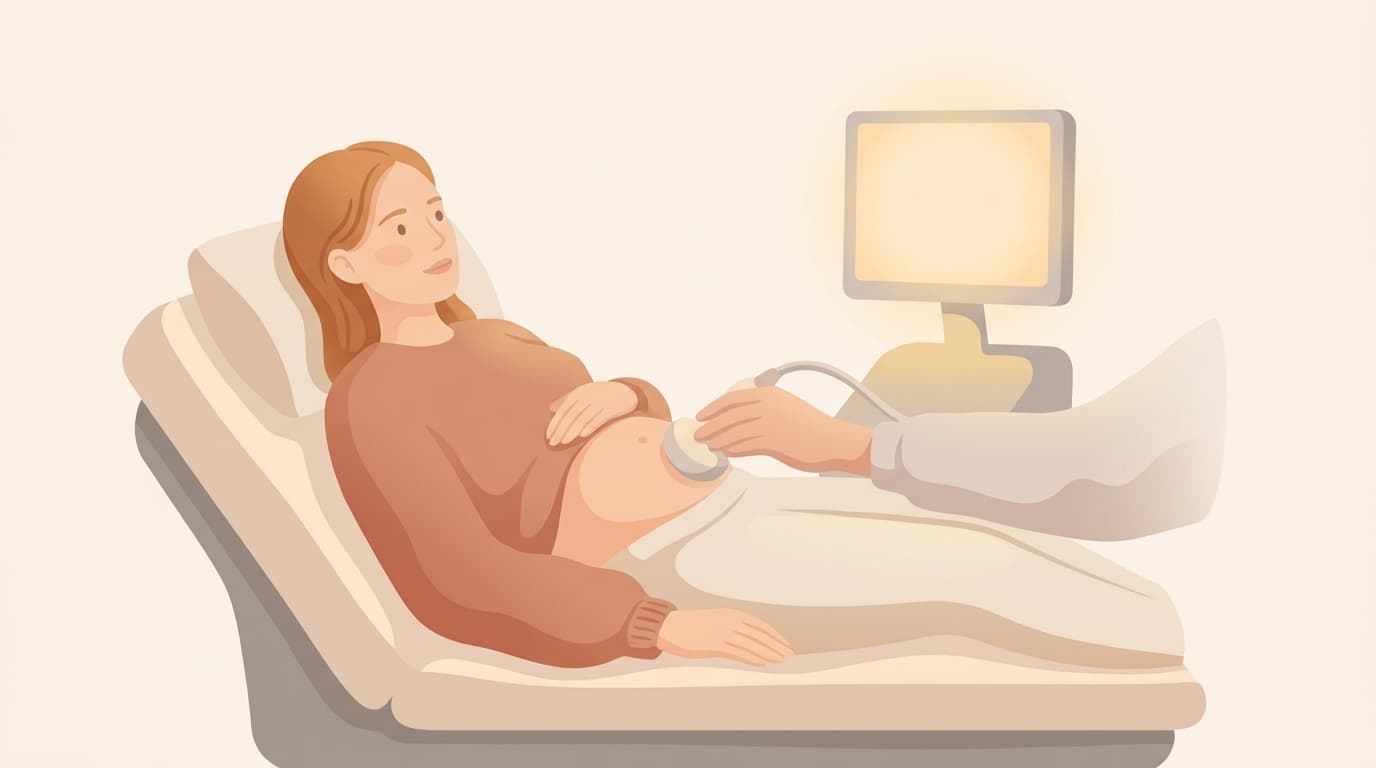
1. Duplex ultrasound (often first). A transabdominal or transvaginal ultrasound with Doppler — ideally with manoeuvres like bearing down (Valsalva) that provoke reflux. It's radiation-free and good at showing pooling and backwards flow, but it depends heavily on the operator knowing what to look for.
2. MR or CT venography. Cross-sectional imaging timed to light up the veins. This maps dilated ovarian veins, pelvic varices and — importantly — the compression points behind May-Thurner and Nutcracker syndromes. If you already have MRI images, a venous-trained radiologist can sometimes re-read them, but only if the study captured the veins at all.
3. Catheter venography (the definitive test). A day procedure where contrast dye is injected directly into the pelvic veins through a thin catheter. It shows exactly which veins leak and how badly, often measures pressures, and can use intravascular ultrasound (IVUS) to confirm compression. It's usually done when treatment is already on the table — diagnosis and treatment planning in one visit.
Which specialist actually does this?
Most women reach a diagnosis through an interventional radiologist or a vascular specialist — the clinicians who both image and treat veins. A gynaecologist is still a sensible first stop, because other causes of pelvic pain (endometriosis among them) need considering too; the key is that venous causes stay on the list rather than falling off it.
What to bring to the appointment
Specialists act on patterns. Before your visit, note:
- When the pain is worst (end of the day? after standing? before your period?)
- What reliably eases it (lying down? legs up?)
- Whether one side dominates
- Any visible veins — vulva, thighs, buttocks — and when they appeared
- Prior scans: where, when, and what type — reports can be re-requested
A KINUUM check produces exactly this kind of doctor-ready summary from your answers, including the questions worth asking.